 Knee Health
The knee joint is the biggest and the most complex joint in the human body which consists of bones, ligaments, tendons, muscles, cartilages, menisci, bursas and the joint capsule. In cases when any of these knee structures are injured human may have severe knee pain and difficulty in walking. When any of knee structures is injured, there may be a sound of popping, sensation of snapping, impassibility, tingling, swelling, limping and immobility of the knee. If you have any of mentioned symptoms, you should visit your doctor for your knee examination to see what is wrong with it and to solve this problem.
The doctor usually begins the knee examination with asking about patient symptoms and history of any injury, not only about those which are connected with knees. When these conditions are clear, doctor starts a physical examination of patient knee. It should be mentioned, that both medical history and physical examination are necessary for proper diagnosis and treatment program. For examination of knee patient is asked to lie or sit down. What is important, doctor examines both knees, comparing injured knee with the healthy one.
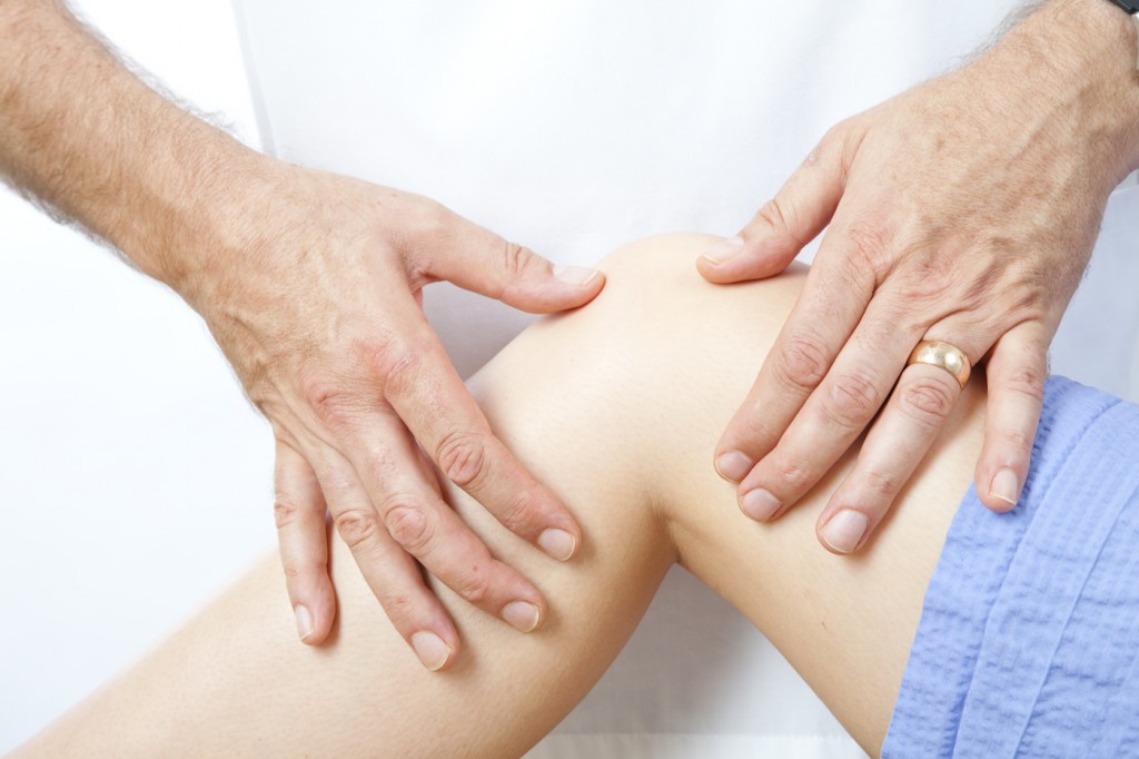
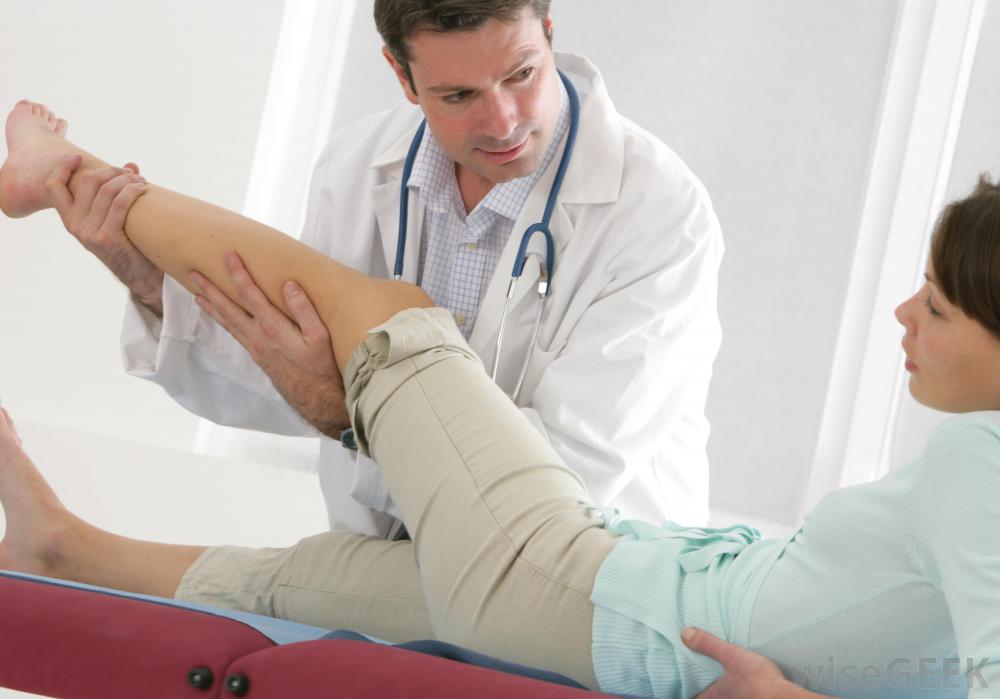
The doctor assesses injured knee for deformity, swelling, redness or any other changes or abnormalities. When observation is done, doctor performs palpation which is the part of knee examination where doctor feels injured knee for temperature, tenderness, swelling, blood flow and some altered sensation. When palpation is done, doctor evaluates knee’s range of motion through active and passive tests. To perform the active test patient is asked to move all joints through a full range of motion, but during passive test joints are moved by the doctor. During these tests doctor listens for any sound of popping, clicking or grinding in joints.
All of following tests are performed to evaluate four ligaments of the knee, namely, valgus and varus tests, posterior drawer test, Lachman test, pivot shift test, McMurray test and arthrometric test.
Valgus and varus tests
These tests are intended for evaluation of medial and lateral collateral ligaments. To perform valgus and varus tests doctor places on hand on the knee joint and other hand on the ankle, and moves patient leg side to side.
Posterior drawer test
This test is meant for evaluation of posterior cruciate ligament. To perform the posterior drawer test the foot is stabilized on the table and the knee is placed at 90 degrees angle. When it is done, the doctor places hands around the knee and pushes the top of the knee with the thumb.
Lanchman test
This test is intended for diagnosis of ACL tear. To perform the Lanchman test the knee is slightly flexed while the patient is laying on its back. The doctor places hand on the tight and pulls the shin to evaluate the softness or firmness of the ligament and to assess any shifting of the shin bone.
Pivot shift test
This test is meant for evaluation of ACL. To perform the pivot shift test the leg has to be fully extended – the doctor holds the ankle with one hand and applies a valgus stress to the knee with other hand, internally rotating the tibia. This test is usually performed after receiving anesthesia and before knee arthroscopy.
McMurray test
This test is intended for diagnosis of meniscus tears. To perform the McMurray test the doctor holds the knee with one hand and the bottom of foot with other hand. When it is done, leg is pushed up while pressing on the knee and turning the leg.
Arthrometric test
This test is meant for those patients who have severe knee pain and thus difficulty to perform an examination. To perform the arthrometric test the doctor uses an instrument which is called an arthrometer – the arthrometer is attached onto lower leg with two sensor pads (one on the patella and other on the tibial tubercle). When the arthrometer is attached, the doctor pushes or pulls on the pressure handle, measuring the pressure.
It should be mentioned, that physical examination tests are not the only options for knee examination – there are a lot of other possibilities as well, for example, knee x-ray, magnetic resonance imaging, arthrocentesis of the knee and arthroscopy. A plain x-ray film of the knee is the best initial imaging test for most knee conditions – the doctor can see most of the knee injuries or disorders without physical examination tests. During magnetic resonance imaging or MRI scan high-energy magnetic waves are used, therefore, the MRI scanner creates highly detailed images of the knee. Thanks to quality of the MRI scan, this is the most-often used method for detecting injuries of ligaments and menisci. Arthrocentesis of the knee, also known as joint aspiration, is used in cases, when patient has severe swelling, because the doctor may find it difficult to examine swollen knee. Arthrocentesis is a procedure in which a needle is inserted into the joint space inside the knee to remove the excess fluid and to look for some infection, inflammation or bleeding. Through this procedure various forms of arthritis may be diagnosed and it may also relieve the pain and make the examination more comfortable. Arthroscopy is a surgical procedure which allows to perform the knee examination, using an endoscope.
Mathew Foster
I am Mathew Foster – an enthusiast of sports who not only regularly practices different sports, but also has a deep interest in it.