 Knee Health
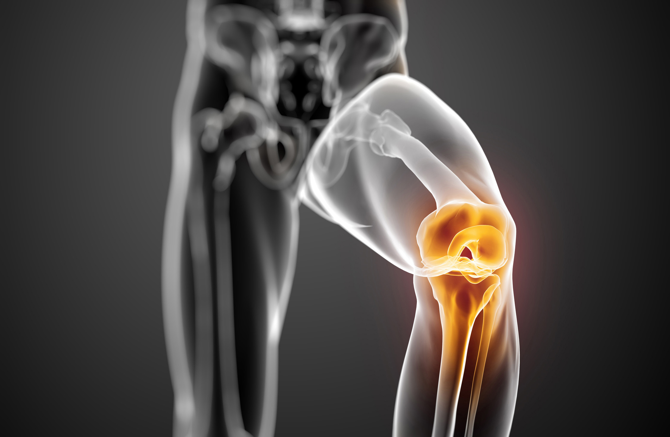
Maybe you have not thought about it, but the knee is the largest and the most complex joint in the human body – it consists of 4 bones and an extensive network of ligaments and muscles. The knee connects the femur (tight bone) with the tibia (shin bone).
Tendons connect knee bones with leg muscles which are moving the knee joint.
Ligaments connect knee bones and are providing stability of the knee. There are four types of ligaments – the anterior cruciate ligament (ACL), the posterior cruciate ligament (PCL), medial collateral ligament (MCL) and the lateral collateral ligament (LCL). Each of them has particular functions in helping to maintain optimal knee stability in a variety of different positions.
- The anterior cruciate ligament or ACL is so called because its form a cross in the middle of the knee joint. ACL reaches from the anterior of the tibia to the posterior of the femur. ACL ensures stability of the femur and the tibia, namely, this ligament prevents the tibia from sliding forward on the femur or the femur from sliding backward on the tibia.
- The posterior cruciate ligament or PCL wraps around the ACL and attaches from the posterior surface of the tibia to the anterior surface of the femur. PCL ensures stability of the femur and the tibia, namely, this ligament prevents the tibial from sliding backward on the femur or the femur from sliding forward on the tibia.
- The medial collateral ligament or MLC is a band which runs between the inner surfaces of the femur and the tibia. It has two parts – an outer part which attaches to the tibia bone and an inner part which attaches to the cartilage in the inside of the knee. MLC ensures stability of the femur, namely, this ligament prevents the femur from sliding side to side, protects the knee from collapsing inwards, and resists forces which are acting from the outer surface of the knee.
- The lateral collateral ligament or LCL is placed on the outside of the knee and connects the outer surfaces of the femur to the top of the fibula. LCL resists impacts which come from inner surface of the knee.
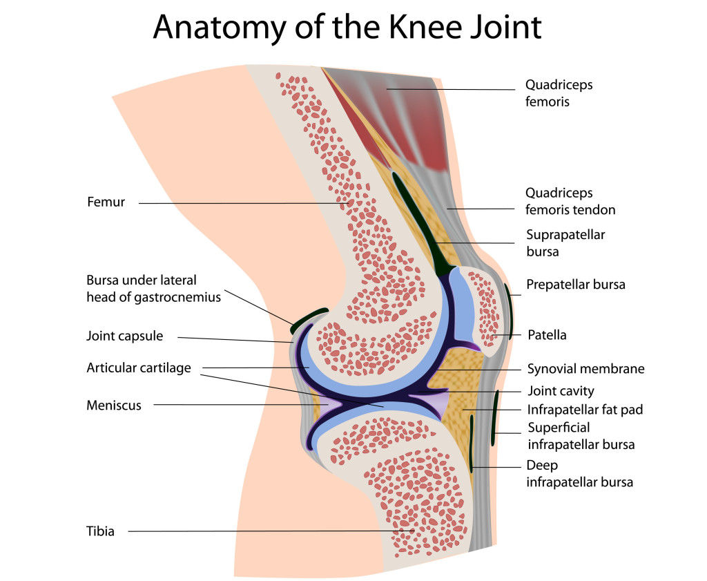
As mentioned before, the knee consists of four main bones – of femur (tight bone), of tibia (shinbone or shankbone), of fibula (outer shin bone) and of patella (kneecap, kneepan). The main movements of the knee joint occur between three of these bones – the femur, patella and tibia.
- The head of the femur articulates with the acetabulum in the pelvic bone, forming the hip joint. The distal part of the femur articulates with the tibia and patella, forming the knee joint. The femur is the strongest and longest bone in the human body. This bone provides possibility of walking and jumping.
- The tibia connects the knee with the ankle bones in the foot. The tibia is found next to the fibula – they are connected by the interosseous membrane of leg, forming a joint which is called a syndemosis. The main function of the tibia is load bearing.
- The outer surface of the tibia is the fibula – a long and thin bone which reaches right down to the ankle joint in the foot. The main function of the fibula is providing of surface for shin muscles to attached to.
- The patella is the knee part which lies in an indentation at the lower end of the femur which is also known as the intercondylar groove. The patella is a thick, circular-triangular bone which articulates with the femur, covers and protects the anterior surface of the knee joint.
Each of these bones is covered with cartilage. The cartilage is an extremely hard, but smooth substance which decreases the frictional forces between bones during movements. Each knee joint has two crescent-shaped menisci which lie on the medial and lateral edges of the top surface of the tibia bone, therefore, these c-shaped pieces of cartilage are called the medial and the lateral menisci. They are some kind of cushioning which ensures shock absorbing between the tibia and the femur and allows for correct weight distribution between these two bones.
The joint capsule is a thick and ligamentous structure which surrounds the entire knee joint. This capsule contains specialized membrane (synovial membrane) which ensures nourishment to all the surrounding structures of the knee. This special membrane produces synovial fluid which lubricates lubricates the knee joint. The capsule is strengthened by surrounding ligaments.
The knee contains numerous bursae (sacs which are filled with fluid) which help the knee to move smoothly and infrapatellar fat pad which functions as cushion against outer forces on the knee.
The knee joint includes two main groups of muscles – quadriceps and hamstrings. Both of these muscles are moving and stabilizing the knee joint.
- The quadriceps consists of four different individual muscles (biceps femoris, vastus medialis, vastus intermedius, vastus lateralis) which form the quadriceps tendon. This tendon connects the muscle with the patella. Contraction of the quadriceps pulls the patella upwards, thereby extending the knee.
-
The hamstring muscles are placed at the back of the tight and they consist of biceps femoris, semitendinosus and semimembranosus. Their functions are flexing and bending of the knee, as well as providing stability of sides of the knee joint.
Mathew Foster
I am Mathew Foster – an enthusiast of sports who not only regularly practices different sports, but also has a deep interest in it.